
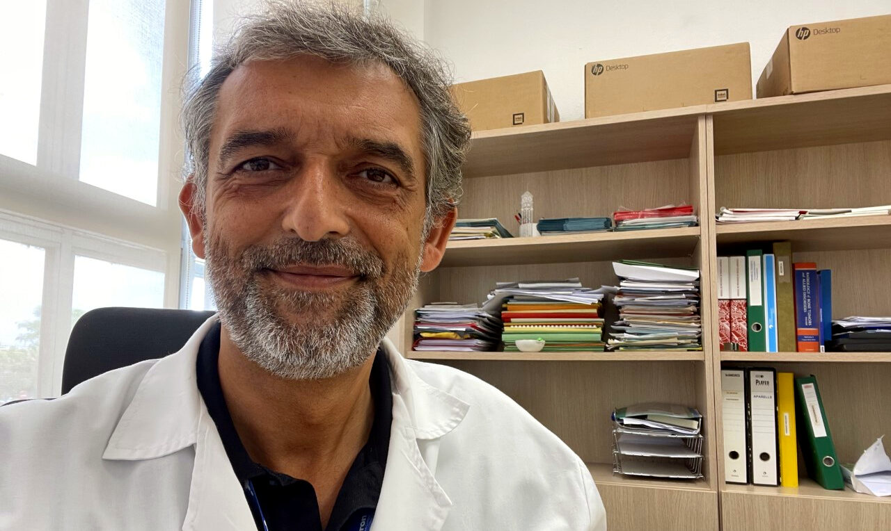
Manel Escobar is currently the Clinical Director of the Diagnostic Imaging Department at Vall d’Hebron University Hospital. He has been a doctor since 1991, and a specialist in Radiology since 1996. He has worked in all imaging modalities, such as Conventional Radiography (CR), Ultrasound (US), Computed Tomography (CT), Magnetic Resonance Imaging (MRI) and hybrid imaging as Positron Emission Tomography with Computed Tomography (PET-CT). He also holds an Executive Master in Healthcare Management. His fields of interest and expertise are Oncologic imaging and Artificial Intelligence applied to medical images.
” I would like to contribute my vision and ideas in order to validate clinical application of supercomputing based algorithms for a new ultrasound method to detect breast cancer. We are facing a revolutionary method of breast cancer detection and characterization, that could complement or even replace mammography in the future, providing a more precise, not using radiation tool, for radiologist and patients”
Why did you choose this profession and what motivated you to do it?
I choose Radiology as a profession because of its unique combination of medical and technical knowledge. During the last five decades, progress in fields as physics, mathematics, informatics and bioengineering have been successfully applied to medical images and radiology, resulting in the more transforming and revolutionary medical speciality.
What will be your contribution to the QUSTom project?
From my position of Clinical Director of Diagnostic Imaging Department at Vall d’Hebron University Hospital, and investigator of Vall d’Hebron Institute of Research and Vall d’Hebron Institute of Oncology, I would like to contribute my vision and ideas in order to validate clinical application of supercomputing based algorithms for a new ultrasound method to detect breast cancer. We are facing a revolutionary method of breast cancer detection and characterization, that could complement or even replace mammography in the future, providing a more precise, not using radiation tool, for radiologist and patients.
Do you think that QUSTom and the solution it proposes can contribute to areas that go beyond cancer? In this sense, what do you hope QUSTom can achieve beyond the life of the project?
In addition to cancer, there are many pathologies that are diagnosed through the use of ultrasound, covering areas as breast, liver, kidney, muscle skeletal system, heart, etc. In this sense, we can think that QUSTom could be used in the future to achieve greater diagnostic precision in different diseases.
I hope QUSTom project will show excellent results in the diagnosis of breast cancer, and could become the new reference method for an earlier and more accurate diagnosis.
How do you like it so far?
Preliminary breast images obtained with the combination of a new ultrasound device and a supercomputer based algorithms are very promising: they are 3D images with an amazing spatial resolution, so it could be possible to detect breast cancer at an initial stage. We have to develop clinical validation of this technology through QUSTom project.
Do you have any advice for young researchers who would like to follow in your footsteps?
I would say: think outside the box! Classical approaches to healthcare problem solving must be questioned. Diagnostic medical images field is probably the one that will develop more in the future, due to the impact of artificial intelligence, machine and deep learning.
