
- BSC coordinates QUSTom, the first project that will use supercomputing to detect tumors more effectively and safely.
- The new imaging modality will improve breast cancer diagnosis and potentially replace mammograms.
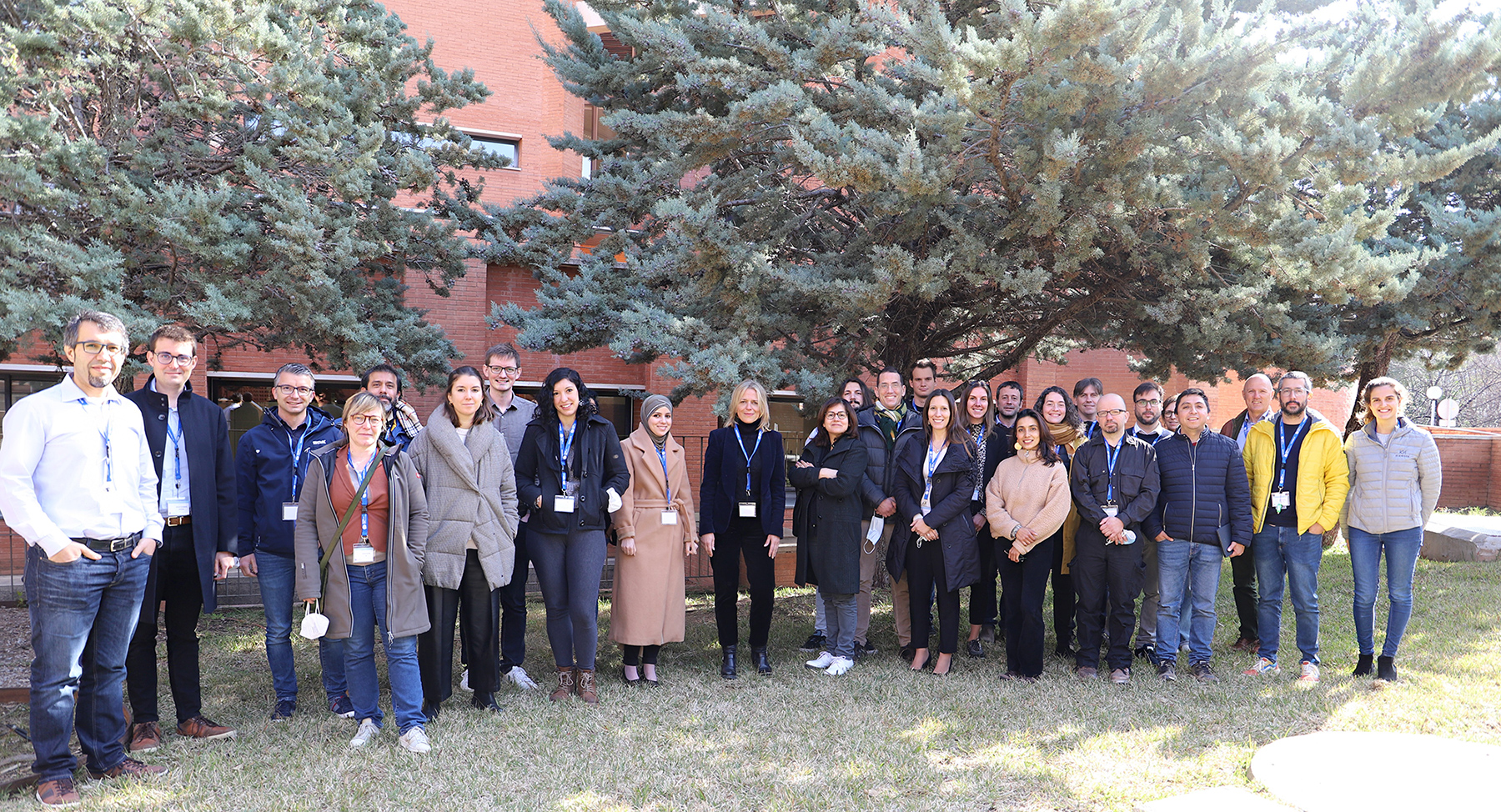
- The consortium brings together physicists, engineers, exploitation experts and radiologists with the aim of applying this technology to patients through the BSC spin-off start-up FrontWave Imaging and Imperial College London.
- QUSTom has been selected to be part of the first call of the Pathfinder Open programme of the European Innovation Council, which aims to support disruptive ideas and projects with great international potential.
The Barcelona Supercomputing Center (BSC) coordinates QUSTom (Quantitative Ultrasound Stochastic Tomography), a new European project that aims to introduce a new medical imaging modality based for the first time on ultrasound and supercomputing, which will complement or even replace current techniques that use X-rays such as mammograms. This technology will be completely safe for patients as it does not use any type of radiation. It will also offer a superior image quality and better monitoring of tumors, among other advantages.
The algorithms that will be developed to obtain the medical images will offer two types of images simultaneously: the image of the patient’s tissue, and the image of its associated uncertainty, which shows, pixel by pixel, how reliable the information is. The project also incorporates concepts such as multimodal imaging and real 3D imaging, which is an unprecedented combination in ultrasound breast imaging.
These algorithms, which will be developed using supercomputers within BSC, will be inspired by others that have proven effective in completely different research areas such as the analysis of the earth’s subsurface.
Apart from BSC, the project has five other partners: Karlsruher Institut für Technologie, Vall d’Hebron Research Institute – VHIR, Arctur and the BSC and Imperial College London spin-off FrontWave Imaging, which is aligned with the objectives of this project, as well as Imperial College London itself as an associate partner. Therefore, physicists, engineers, operational experts and radiologists will work together to develop the next generation of radiation-free, accurate and scalable breast cancer diagnostic tools.
BSC researcher and project coordinator Josep de la Puente says: “QUSTom poses an excellent opportunity to bring ultrasound imaging to the next level. Interestingly enough, the revolution that we propose comes, not just from an extraordinary imaging device, but from the imaging algorithms used to generate unprecedented ultrasound images. Images that we can fairly compare to those obtained with MRI”.
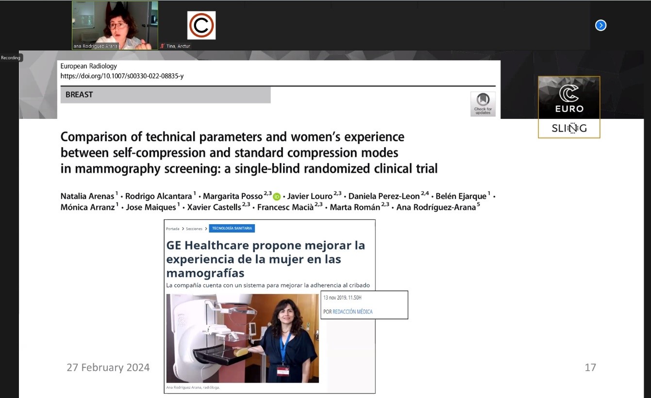
The process of understanding, interpreting and configuring the images as a new diagnostic tool will be carried out by the team of Breast Imaging Radiologists from the Women’s Radiology Service at Vall d’Hebron Hospital. “The development of this new technology will be carried out within the framework of the multimodal assessment that the team performs in the clinical care process as an integral part of the diagnosis and monitoring of breast cancer, all in the context of the Breast Pathology Unit,” highlights Ana Rodríguez-Arana, head of the Women’s Radiology Service at the Vall d’Hebron Hospital.
QUSTom has been selected to take part in the first call of the Pathfinder Open programme of the European Innovation Council (EIC), funded by the European Union’s Horizon Europe Framework Programme, which aims to support disruptive ideas and projects with great international potential. The project has been awarded with 2,744,300 euros over 2 years. In this first call the European Commission evaluated a total of 868 projects and in the end only 56 were selected, 11 of them from Spain. In total, they will receive up to 168 million euros in funding.
Ultrasound to revolutionize diagnosis of the most common tumor
Breast cancer is the most frequently diagnosed type of tumor in the world, with 2.3 million women diagnosed in 2020 (REf SEOM) and 700,000 deaths due to this disease that same year. Early detection is therefore essential, since, if successful, survival at 5 years after diagnosis is as high as 90%.
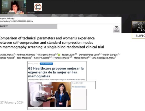
Mammography is one of the most widely used methods to detect breast cancer and has saved millions of lives. However, there are studies that claim that it can give false positives and alert of a possible tumor that is not found later in the screening phase.
“We are very ambitious and plan for validation of the technology within the project’s lifetime. We are also working on a roadmap towards its actual exploitation, we don’t want this technology sitting in the lab. We are leaving no stone unturned towards satisfying what is, in our eyes, an urgent need for the female population worldwide. The challenge is huge but all of our partners and associates are extraordinarily committed and motivated towards our mission”, concludes de la Puente.
